Medical progress often unfolds through small, steady steps rather than dramatic breakthroughs. That’s why staying up to date is essential: by researching, reading, discussing, and learning from one another. From June 12th to 15th, 2024, we answered the call of EULAR (European Alliance of Associations for Rheumatology) and headed to Vienna.
Under the Austrian patronage of Daniel Aletaha, rheumatologist and current president of EULAR, the annual congress took place. Let us share with you our top highlights from EULAR 2024.

EULAR is one of the most important platforms for scientific and clinical exchange in Europe, aimed at improving the lives of people with rheumatic and musculoskeletal diseases. Besides rheumatology, radiology plays a crucial role in diagnosis and treatment selection.
Technical advancements in imaging often lead to significant changes, including shifts in terminology. One example is the new classification of Bechterew’s disease into:
The new term axSpA also encompasses early forms of the disease, before the spine shows signs of ossification (ankylosing spondylitis).
A similar issue has been a topic of discussion for some time among professional societies focused on axial spondyloarthritis (ASAS—Assessment of SpondyloArthritis international Society) and psoriatic arthritis (GRAPPA—Group for Research and Assessment of Psoriasis and Psoriatic Arthritis): Is it axSpA or axPsA?
One EULAR session evolved around the question: How can we distinguish between axial spondyloarthritis and axial psoriatic arthritis, and what commonalities exist? Could they possibly be the same disease with different phenotypes?
This intriguing session was chaired by rheumatologist Dennis McGonagle from Leeds, United Kingdom, and rheumatologist Gizem Ayan from Ankara, Türkiye. Rheumatologist Phillippe Carron from Ghent, Belgium, provided a comprehensive overview of axial involvement patterns. He highlighted some similarities between axPsA and axSpA through imaging findings, but also pointed out distinct differences in the lesions.
Rheumatologist Sofia Ramiro traveled from Leiden, Netherlands, to present current results from clinical research. She emphasized the need for further studies, particularly to explore the efficacy of biologics such as interleukin-23 inhibitors. Similarly, rheumatologist Catarina Abreu from Lisbon presented various phenotypes of axPsA and concluded by stressing the importance of refining the classification criteria for axPsA.
This is where current research comes into play—highlighted by the AXIS study presented by rheumatologist Murat Torgutalp from Charité hospital, Berlin, Germany. Specialists from the BerlinCaseViewer team are also involved in this study, including Torsten Diekhoff (Radiology), Kay G. Hermann (Radiology), and Denis Poddubnyy (Rheumatology).
Preliminary study findings are indeed interesting: Axial psoriatic arthritis often goes hand in hand with elevated levels of C-reactive protein (CRP) and positive results for HLA-B27, alongside back pain, often linked with inflammatory and structural lesions in the sacroiliac joints and along the spine. Such changes can also be detected using imaging techniques.
Participants keen to experience the synergy of rheumatology and radiology in action were well served by interactive sessions supported by the BerlinCaseViewer app.

Our first interactive session on June 13th, 2024 was a resounding success: the small room was filled to capacity, and there was a long waiting list. This did not dampen the enthusiasm for learning; quite the contrary! Equipped with laptops on-site and the BerlinCaseViewer app in hand, participants could analyse cases close-up with imaging findings in pixel-perfect quality.
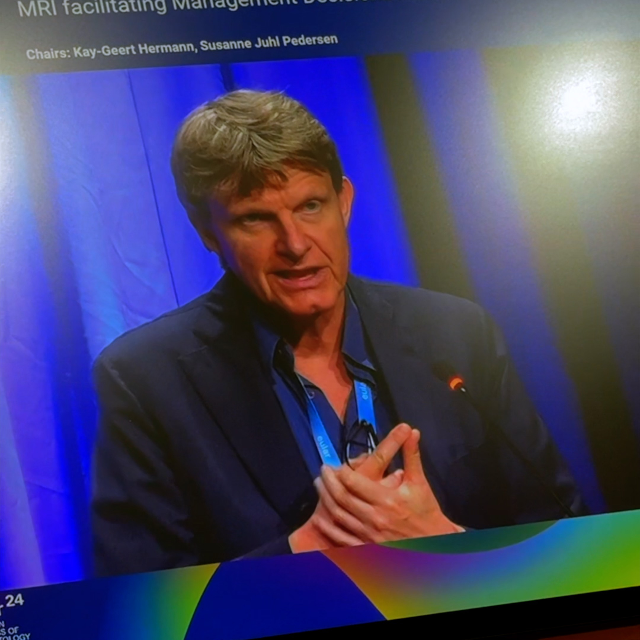
Radiologist Kay G. Hermann from the BCV team co-hosted the session on axial spondyloarthritis with Belgian rheumatologist Gaëlle Varkas.
At a Glance:
During interactive case work, participants had the opportunity to examine and evaluate typical findings of axSpA from X-rays and MRI scans. The cases at hand, with differential diagnoses included, sparked lively discussions, underscoring the importance of integrating theoretical knowledge with practical application.
Those working in rheumatology or musculoskeletal radiology understand that rheumatic diseases can affect not only the axial skeleton but the entire body. Hence, in our second interactive session on June 14th, 2024, we focussed on extremities.
Susanne Juhl Pedersen (Rheumatology) and Kay G. Hermann (Radiology) led the session in a larger room this time. Participants brought their own devices, with the BerlinCaseViewer platform at their fingertips to interactively work on cases—no lengthy installation needed.
At a Glance:
After a brief theoretical introduction, we dove straight into practical demonstrations to showcase how magnetic resonance imaging can assist in diagnosing and managing rheumatic musculoskeletal disorders. Following the session, the speakers were available through the Pin & Comment feature to answer questions about the cases, including those that couldn’t be fully discussed due to time constraints.
You can explore one of these cases through our web app. A 63-year-old male presents with a medical history notable for pain in the metacarpophalangeal (MCP) joints without any accompanying swelling, and morning stiffness lasting approximately 30 minutes. Clinical findings reveal that he is positive for both anti-citrullinated protein antibodies (ACPA) and rheumatoid factor (RF). His C-reactive protein (CRP) level is measured at 3.5 mg/l. What condition are we dealing with here?
Taking these the MRI findings into account, can you steer the diagnosis in the right direction?
EULAR 2024 provided numerous new insights with the latest research findings. Besides the interactive sessions with the BerlinCaseViewer, the comparison between axSpA and axPsA was particularly enlightening for us.
The BCV app already includes several modules on axial spondyloarthritis (axSpA), serving as an outstanding resource for professionals looking to expand their specialized knowledge. For us, it underscores that axPsA also offers potential for exciting modules.
Stay tuned for future developments and new modules: Gain fresh expertise with the BCV Insider, our newsletter, or follow us on Instagram or LinkedIn for current insights.
Stay Updated and subscribe to our Newsletter.
Signup for news and special offers!
You can unsubscribe anytime. For more details, review our Privacy Policy.


